Latent Tuberculosis Infection

SPOT the hidden DANGER of latent TB


Active TB vs Latent TB

Of the global population, approximately 2 billion people is estimated to be infected with Mycobacterium tuberculosis1. There are two types of tuberculosis (TB) conditions: active tuberculosis (TB) disease and latent TB infection (LTBI). If untreated, approximately 5%–10% of persons with LTBI may progress to active TB disease, passing the infection to others2 According to the WHO End TB Strategy, systematically providing TB preventive treatment to those at highest risk of developing active TB will prevent the development of disease and reduce the risk of transmission in the population; this is a critical step to End TB.

Latent tuberculosis infection (LTBI) is a state of persistent immune response to stimulation by M. tuberculosis antigens without evidence of clinically manifested active TB3. The immune system cannot eliminate TB bacteria completely instead it is contained in the granuloma. They may develop disease in the future when the immune system weakens, making the person ill and at risk of spreading the infection.


The WHO’s annual report4 on the status of global efforts to End TB notes that, at the present trajectory, most WHO regions and individual countries will fall significantly short of the End TB milestones. Hence, global TB prevention efforts along with targeted testing and treatment of latent TB infection builds a strong core TB infrastructure to realizing TB elimination. For an infection that kills 1.3 million a year, there’s routine screening or a gold standard test for LTBI5 – Either a tuberculin skin test (TST) or interferon-gamma release assay (IGRA) is used. A diagnosis of LTBI first requires that active TB disease be excluded by medical evaluation.

IGRAs are whole-blood tests that measure the individual’s immune reactivity to tuberculosis. Leukocytes from most individual who have been infected with M. tuberculosis will release interferon-gamma (IFN-g) when mixed with antigens derived from M. tuberculosis. To conduct the test, fresh blood samples are extracted and mixed with antigens and controls. Two IGRAs approved by the U.S. Food and Drug Administration (FDA) are6:

  • QuantiFERON® – TB Gold In-Tube test (QFT–GIT);
  • SPOT® TB test (T–SPOT.TB)

The antigens, testing methods, and interpretation criteria for IGRAs differ as below:


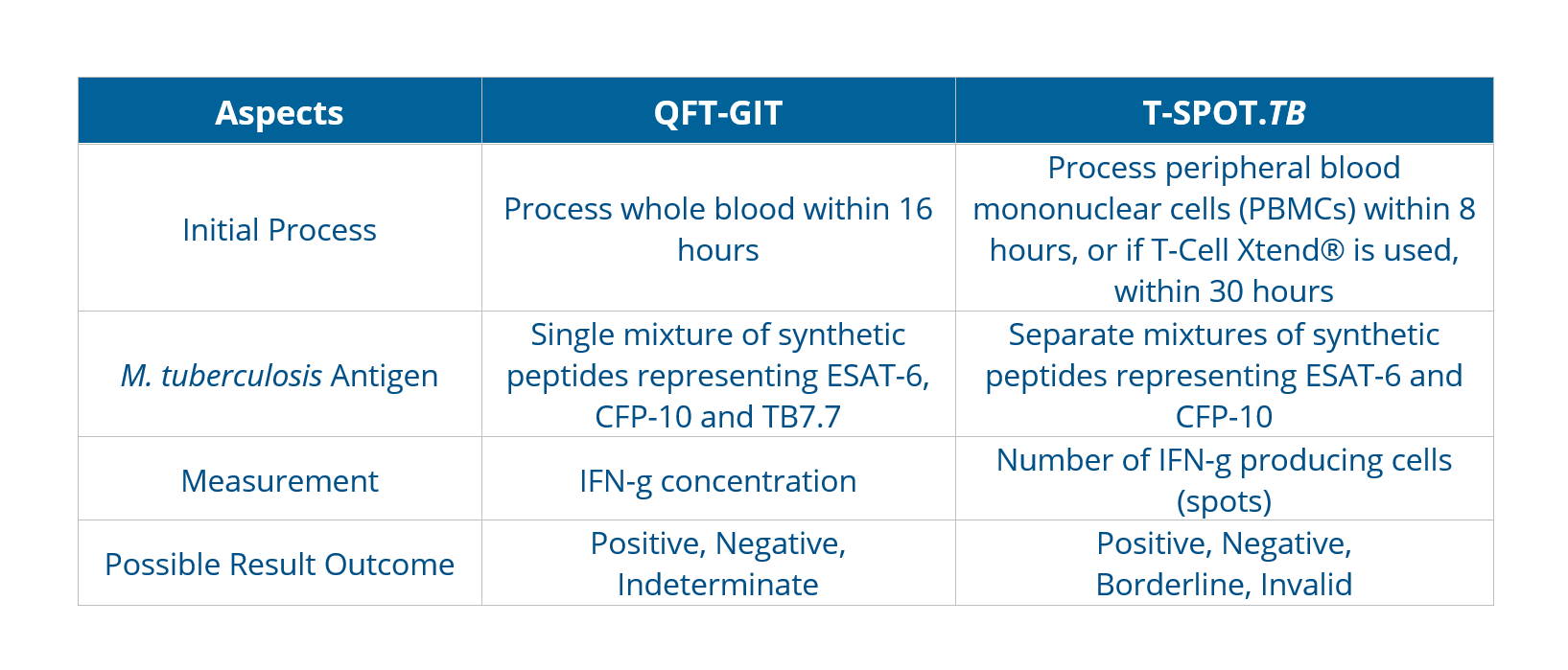
The incremental cost-effectiveness of IGRAs and TSTs appears to be influenced mainly by their accuracy. Bacille Calmette-Guérin (BCG) vaccination plays a decisive role in reducing the specificity of TST, leading the choice towards adopting IGRA-only strategies for better diagnostic accuracy.As with TSTs, IGRAs should be used as an aid in diagnosing latent infection with M. tuberculosis. A positive test result suggests that M. tuberculosis infection is likely; negative result suggests that infection is unlikely. An indeterminate result or borderline test result (T-Spot only) indicates an uncertain likelihood of M. tuberculosis infection6.

NOT all IGRAs are the same.

The T-SPOT.TB test is the only globally regulated IGRA that is normalised for both cell number and culture conditions in each patient specimen. The cell enumeration technology in the proprietary T-SPOT.TB test removes serum factors that could adversely affect the test result, making it the most sensitive and most specific test for TB infection7, supported by clinical data obtained even in challenging patient populations.

Learn more about the T-SPOT.TB Test here.


References:

  1. Houben RM, Dodd PJ. The global burden of latent tuberculosis infection: a re-estimation using mathematical modelling. PLoS Med 2016;13:e1002152.
  2. Sutherland I. Recent studies in the epidemiology of tuberculosis, based on the risk of being infected with tubercle bacilli. Adv Tuberc Res 1976;19:1–63.
  3. Latent tuberculosis infection: updated and consolidated guidelines for programmatic management. Geneva: World Health Organization; 2018.
  4. WHO End TB Strategy. World Health Organization. WHO End TB Strategy. Published September 8, 2015. Assessed January 9, 2020.
  5. WHO End TB Strategy. Latent tuberculosis infection Updated and consolidated guidelines for programmatic management. Published 2017, Assessed November 30, 2021 via https://apps.who.int/iris/bitstream/handle/10665/260233/9789241550239-eng.pdf
  6. Centres for Disease Control and Prevention, Interferon-Gamma Release Assays (IGRAs) – Blood Tests for TB Infection Fact Sheets. Published May 4, 2016. Assessed November 30, 2021 via https://www.cdc.gov/tb/publications/factsheets/testing/igra.htm
  7. Oxford Immunotec. T-SPOT.TB Package Insert PI-TB-IVD-UK V3. Abingdon, UK. February 2019
Share this article

Abionic Early Sepsis Detection with Pancreatic Stone Protein (PSP)

Early Sepsis Detection with Pancreatic Stone Protein (PSP)

Sepsis

Sepsis is defined as life-threatening organ dysfunction caused by a dysregulated host response to infection.

Early recognition of sepsis is essential and is a major determinant of the disease’s outcome, according to The WHO and the Surviving Sepsis Campaign.

However this has proven to be challenging as the confirmation of sepsis diagnosis is based largely on nonspecific clinical signs, laboratory findings and medical scores which are usually obtained after sepsis onset.

Currently, C-reactive protein (CRP) and procalcitonin (PCT) are biomarkers routinely used for patients suspected of sepsis. Pancreatic stone protein or PSP is an emerging biomarker showing promise as it is characterized by higher accuracy in the diagnosis and prognosis of sepsis compared to CRP and PCT.

The Pancreatic Stone Protein (PSP)

• A 16 kDa polypeptide of the C-type lectin family of protein.
• Mostly secreted by the acinar cells of the pancreas but also by the intestine and stomach.
• An early sensor of sepsis and multiple organ dysfunction acting as an “alert signal” to help clinicians provide adequate infection control strategies and organ support to restore homeostasis.

Performance

PSP was proven to be more accurate, with a higher sensitivity, specificity, positive and negative predictive values than CRP, PCT, IL-6 and other cytokines for the diagnosis of sepsis and prognosis of unfavourable outcomes in multiple studies and clinical settings (ED, ICU, surgical, nonsurgical adult and children).

It also performed well in studies involving a variety of critically ill patients including severe burns, polytrauma, post-cardiac surgery and on admission to the ICU.

Unique Characteristic

PSP may start to increase above the normal level before the development of clinical signs and symptoms of sepsis.

It was found to be the only biomarker able to identify sepsis 72 hours before clinical diagnosis, thereby providing a large window of opportunity for timely initiation of accurate clinical management.

PSP Measurement

Can now be done via a Point-of-Care(POC) device, which is uncommon for CRP and PCT in ICU, leading the way for simple, on-demand, around-the-clock, serial biomarker assessments instead of one-off testing upon clinical suspicion of sepsis.

This is key to faster treatment decisions, reducing mortality and lowering sepsis-related healthcare costs.

The IVD CAPSULE PSP on the abioSCOPE® is the first CE-marked in vitro diagnostic test to enable fast, reliable and early sepsis detection at the pointof-care from a single drop of blood in only 5 minutes.

Click the link below for more information:

Products | Abionic


References:

Eggimann, Philippe & Que, Yok-Ai & Rebeaud, Fabien. (2019). Measurement of pancreatic stone protein in the identification and management of sepsis. Biomarkers in Medicine. 13. 10.2217/bmm-2018-0194.

Pugin, J., Daix, T., Pagani, JL. et al. Serial measurement of pancreatic stone protein for the early detection of sepsis in intensive care unit patients: a prospective multicentric study. Crit Care 25, 151 (2021). https://doi.org/10.1186/s13054-021-03576-8

World Health Organization (WHO). Sepsis. https://www.who.int/news-room/fact-sheets/detail/sepsis

Rudd KE, Johnson SC, Agesa KM, Shackelford KA, Tsoi D, Kievlan DR, et al. Global, regional, and national sepsis incidence and mortality, 1990-2017: analysis for the Global Burden of Disease Study. Lancet (London, England). 2020;395(10219):200-11.

Klein HJ, Niggemann P, Buehler PK, Lehner F, Schweizer R, Rittirsch D, Fuchs N, Waldner M, Steiger P, Giovanoli P, Reding T, Graf R, Plock JA. Pancreatic Stone Protein Predicts Sepsis in Severely Burned Patients Irrespective of Trauma Severity: A Monocentric Observational Study. Ann Surg. 2021 Dec 1;274(6):e1179-e1186. doi: 10.1097/SLA.0000000000003784. PMID: 31972652.

Keel et al., 2009 and Reding et al., 2018 Pathophysiological mechanisms of PSP function in Sepsis- Abionic slides; Abionic brochure

Share this article

Medical Solutions

Digital health promises to change the face of healthcare. Reflecting this is growing interest in the digital health space, as evidenced by a substantial $4.7 billion being invested in 2017, as outlined in the brand new report from IDTechEx Research, Digital Health 2018: Trends, Opportunities and Outlook. Digital health also took prominence in the JP Morgan Healthcare Conference in January 2018, illustrating its growing importance – but it is not limited to strictly the healthcare space. CES 2018 also saw digital health having a substantial presence marking a new age of patient-centric healthcare.

The Rise of Digital Health

Digital health is a convoluted and complex field, much of which is made up of technologies and services that enable healthcare outside of traditional clinical settings. It follows a global trend in the healthcare industry of decentralization to alleviate overburdened hospitals and clinics. Coupled with escalating healthcare costs, shrinking profit margins and ageing populations suffering with chronic conditions, digital health offers a solution to these problems for all players in the space including patients, providers and payers.

In fact, it presents such an alluring and lucrative opportunity that companies not previously in the healthcare space are making significant investments and moves to do so. Big tech companies such as Amazon, Apple and Alphabet also used January 2018 to announce their endeavours into the digital health space, impacting the price of stocks in the healthcare market.

Numerous Factors Encouraging the Rise of Digital Health

The time is ripe for digital health due to the combination of a number of factors. These include changing population demographics, such as ageing populations with increasing prevalence of chronic diseases, as well as current and upcoming changes to regulations and reimbursements which mean that the route to market and take-up of digital health services and technologies is more likely in 2018 and beyond.

The Wide Scope of Digital Health

The IDTechEx Research report Digital Health 2018: Trends, Opportunities and Outlook acts as a primer to the digital health space, providing a detailed overview of the ecosystem and offering insights into the key trends, opportunities and outlooks for all aspects of digital health. The time is ripe for digital health due to the combination of a number of factors. These include changing population demographics, such as ageing populations with increasing prevalence of chronic diseases, as well as current and upcoming changes to regulations and reimbursements which mean that the route to market and take-up of digital health services and technologies is more likely in 2018 and beyond. The IDTechEx Research report Digital Health 2018: Trends, Opportunities and Outlook acts as a primer to the digital health space, providing a detailed overview of the ecosystem and offering insights into the key trends, opportunities and outlooks for all aspects of digital health.

Share this article

Digital Health Emerging in Healthcare


Digital health promises to change the face of healthcare. Reflecting this is growing interest in the digital health space, as evidenced by a substantial $4.7 billion being invested in 2017, as outlined in the brand new report from IDTechEx Research, Digital Health 2018: Trends, Opportunities and Outlook. Digital health also took prominence in the JP Morgan Healthcare Conference in January 2018, illustrating its growing importance – but it is not limited to strictly the healthcare space. CES 2018 also saw digital health having a substantial presence marking a new age of patient-centric healthcare.

The Rise of Digital Health

Digital health is a convoluted and complex field, much of which is made up of technologies and services that enable healthcare outside of traditional clinical settings. It follows a global trend in the healthcare industry of decentralization to alleviate overburdened hospitals and clinics. Coupled with escalating healthcare costs, shrinking profit margins and ageing populations suffering with chronic conditions, digital health offers a solution to these problems for all players in the space including patients, providers and payers.

In fact, it presents such an alluring and lucrative opportunity that companies not previously in the healthcare space are making significant investments and moves to do so. Big tech companies such as Amazon, Apple and Alphabet also used January 2018 to announce their endeavours into the digital health space, impacting the price of stocks in the healthcare market.

Numerous Factors Encouraging the Rise of Digital Health

The time is ripe for digital health due to the combination of a number of factors. These include changing population demographics, such as ageing populations with increasing prevalence of chronic diseases, as well as current and upcoming changes to regulations and reimbursements which mean that the route to market and take-up of digital health services and technologies is more likely in 2018 and beyond.

The Wide Scope of Digital Health

The IDTechEx Research report Digital Health 2018: Trends, Opportunities and Outlook acts as a primer to the digital health space, providing a detailed overview of the ecosystem and offering insights into the key trends, opportunities and outlooks for all aspects of digital health, including:

> Telehealth and telemedicine: the provision of remote clinical or non-clinical services such as doctors visits through video calls e.g. Remote digital examinations for cardiac conditions through measuring heart rate and blood pressure and having a virtual physician meeting.

> Remote patient monitoring: the remote monitoring of individuals to collect information through wearable sensors outside of a clinical settings e.g. smart watches collecting information on blood pressure.

> Digital therapeutics / digiceuticals / software-as-a-drug: using software such as mobile phone apps to diagnose, treat and monitor patients e.g. a mobile app prescribed to treat PTSD.

> Consumer genetic testing: describes the use of genome sequencing on consumer samples to reveal details about an individuals’ genetic make-up which may impact their health e.g. using a cheek swab to determine whether you are likely to be predisposed to particular vitamin deficiencies or whether you are potentially lactose intolerant.

> Diabetes management: chronic diseases such as diabetes present a substantial market for digital health e.g. continuous or flash glucose monitors which are wearable or non-invasive.

> Smart home as a carer: lower costs and sizes of sensors, and better connectivity and networks along with ageing and diseased populations mean that fitting sensors into the home is becoming a reality allowing decentralized healthcare e.g. sensors which alert carers in case of an emergency.

Read more at: https://www.globalbiotechinsights.com/articles/14038/digital-health-is-becoming-a-disruptive-force-in-healthcare

Share this article

Tummy Tech Tracks Electrical Activity for Signs of Indigestion

A new wearable device that non-invasively monitors electrical activity in the stomach could help people with digestive problems determine with greater precision whether treatments or diets are working.

If clinically validated, the stomach sensor could also help revive a medical technology called electrogastrography (EGG) that once piqued gastroenterologists’ interest, but has largely fallen out of favor owing to controversy surrounding its diagnostic relevance.

EGG works much like an electrocardiogram, except instead of recording electrical activity of the heart it picks up electrical signals that travel through stomach muscles to control gastric contractions.

The method is designed to test whether so-called slow waves originating from pacemaker cells of the stomach are oscillating at a healthy 3 cycles per minute. But the signals detected by adhesive electrodes on the skin are often weak and muddied by artifacts of stomach movement.

The technique had a brief clinical heyday in the 1990s, but the world’s gastroenterologists have since largely abandoned it. “I view it as the Rodney Dangerfield of GI motility,” says Abell. “It gets no respect.” 3CPM, one of the only companies with an EGG system still on the market today, sold fewer than 10 units per year in the late-2000s, according to company CEO Mark Noar (although he notes that sales have picked up again, with 45 of the $25,000 machines purchased by customers around the world in 2017).

Todd Coleman and Armen Gharibans, bioengineers from the University of California, San Diego, set out to fix what they saw as pitfalls in the gastric gauging technology. First, they upped the number of channel recordings from the single waveform found in conventional EGGs like those sold by 3CPM to a whopping 25 data-collecting channels. They also created a smaller version with just eight channels to enable mobile recordings on the go.

The researchers next optimized the electrode placement, arranging the dime-sized cups in a grid below the sternum and over the bellybutton. And most importantly perhaps, they developed an algorithm that removes electrical noise (originating from external motion) from the true slow-wave signal associated with internal stomach muscle movements. The algorithm also weights the cleaned-up signals from each electrode to emphasize those relaying the most physiologically relevant readouts.

Coleman and Gharibans teamed up with their UCSD colleague Hayat Mousa, a pediatric gastroenterologist at Rady Children’s Hospital-San Diego, to test the device on 11 kids, aged 7 to 17, each of whom had chronic stomach problems. They affixed their “high-resolution” EGG set-up to each child, while simultaneously administering a more invasive diagnostic procedure called manometry, in which a catheter containing pressure transducers is passed through the nose or mouth to measure contractions at the base of the stomach.

As reported last week in the journal Scientific Reports, the results of the two tests were significantly correlated for each and every youngster evaluated. By comparison, conventional one-channel EGG recordings without the signal-to-noise filtering matched the manometry readings in only three of the 11 study subjects.

“That’s a notable finding,” says David Levinthal, director of the Neurogastroenterology and Motility Center at the University of Pittsburgh. “This is clearly better than what we had before.”

Still, that doesn’t necessarily make the test clinically meaningful.

For one thing, the results indicate that high-res EGG can capture about one-third of the variation in stomach contractions, as measured by manometry—which still leaves the bulk of what’s causing those contractions unexplained.

Plus, the many channels that feed into the EGG setup are mainly used to find the best signal of electrical activity, not the spatial arrangement. That may be valuable for knowing if the frequency of slow wave rhythms is faster or slower than the normal 3 cycles per minute, says Aydin Farajidavar, a biomedical engineer at New York Institute of Technology, “but it doesn’t tell us anything about the pattern distribution of slow wave propagation.”

Moreover, “the stomach is not just a tube that has this wave of contraction going from the esophagus down to the duodenum,” notes Levinthal. “It is a regionally specialized capacitance organ and a mixer and a grinder”—all activities that could be independently disrupted in various stomach diseases.

That leads some critics to dismiss the idea that EGG—even newer and improved EGG methods—will ever have diagnostic value. “Spontaneous EGG by itself has objective physiological limitations that render it unreliable, no matter what,” says Martin Mintchev, a biomedical engineer at the University of Calgary.

Levinthal, however, is keeping an open mind: “It’s worth entertaining the idea that there could be electrical signatures of different disorders,” he says.

Coleman and Gharibans hope to win over skeptics with further experimental data. Already, in collaboration with UCSD gastroenterologist David Kunkel, they’ve tested their prototype on around 25 adults with digestive disorders at the university’s Jacobs Medical Center, and found in as-yet unpublished work that the electrical signals sync with problems such as abdominal pain, heartburn, and bloating. “It actually does correlate with symptom severity,” Coleman says.

Meanwhile, as the hospital-based tests were going on, Gharibans was also hard at work developing an app for keeping tabs on meals, snacks, bowel movements, nausea, vomiting, or any other events of gastrointestinal interest. He also filled a drab-gray, 3D-printed plastic box with an accelerometer, lithium polymer battery, microSD card, and circuit board to record movement patterns and gastric electrical signals from the comfort of his home.

On eight non-consecutive days over the span of about six months, Gharibans strapped the minidisc player–sized device to his own belly for 24-hour-long recording sessions. “You don’t even really notice it too much,” he says. “As long as you don’t sleep on your stomach, it’s fine.”

Gharibans’ measurements revealed how daily activities like sleep, meals, and exercise all impacted the electrical signals in his stomach. He and Coleman now hope to get the portable device into the hands—or onto the bellies—of more patients for at-home monitoring of stomach ailments. Their vision is for people with perpetual stomach issues to wear the high-res EGG and then track in real-time, in a quantitative and objective way, whether any changes in diet or medications are bringing about gastrointestinal relief. “That’s the potential,” Coleman says.

One of the first people (other than Gharibans) to have given the device a try is Dana Lewis, founder of the Open Artificial Pancreas System project, an effort among DIYers with type 1 diabetes to create their own open-source technologies for automating blood-sugar management.

Most evenings, for the past few weeks, Lewis has taken a shower and then stuck a dozen pieces of medical adhesive to her abdomen. One is for her insulin pump, another for her glucose monitor—the two devices needed for Lewis to control her diabetes. The other 10 hold the electrodes for the UCSD team’s high-res EGG.

Lewis hopes the data she gathers about her own stomach movements following different types of meals, when matched to her blood-sugar levels, will help her improve the algorithm behind her closed-loop insulin dosing system.

“Having this picture of stomach activity and digestion will help clear away some of the noise,” she says. “Hopefully we can adapt what we’re doing to match what’s actually happening inside the body.”

Read more here.

Slug Slime Inspires a New Type of Surgical Glue

For a glue that holds up inside the body, turn to the humble slug, Arion subfuscus. A new super-sticky material mimics slug slime’s ability to stick on slick wet surfaces and could lead to more effective medical adhesives.

The material has two parts: a sticky layer that attaches to a surface, and a shock-absorbing layer that reduces strain. That makes the adhesive less likely to snap off.

Researchers tested the material as a surgical adhesive in a number of different scenarios: It stuck to pig skin and liver. It latched on to a beating pig’s heart, even when the surface was coated in blood. It sealed up a heart defect, preventing liquid from leaking even when the organ was inflated and deflated tens of thousands of times. And it was less toxic in the body than a commonly used commercialized tissue adhesive, researchers report July 28 in Science.

The researchers hope the material could someday be used in surgical procedures in place of invasive sutures and staples.

Watch the video here.